Acinetobacter sp. Ver3 encodes two putative SOD enzymes
The genome of Acinetobacter sp. Ver3 (accession quantity JFYL01 21) accommodates two putative SOD genes21. CL42_08295 is positioned on contig JFYL01000023.1, is the second gene inside a putative two-gene operon and displays homology to sodB genes of gram-negative micro organism (Fig. 1). Alternatively, CL42_01680 is positioned on contig JFYL01000003.1, has homology to sodC genes of gram-negative micro organism and is predicted to represent a monocistronic transcription unit.
Surroundings of sod genes in Acinetobacter sp. Ver3. Schematic illustration of the Acinetobacter sp. Ver3 sodB (up) and sodC (down) loci, current in contigs JFYL01000023 and JFYL01000003, respectively. sod genes are proven in purple whereas proximal genes are displayed in purple. The locus tag is offered beneath every gene. Three tRNAs (inexperienced) are encoded within the sodB area.
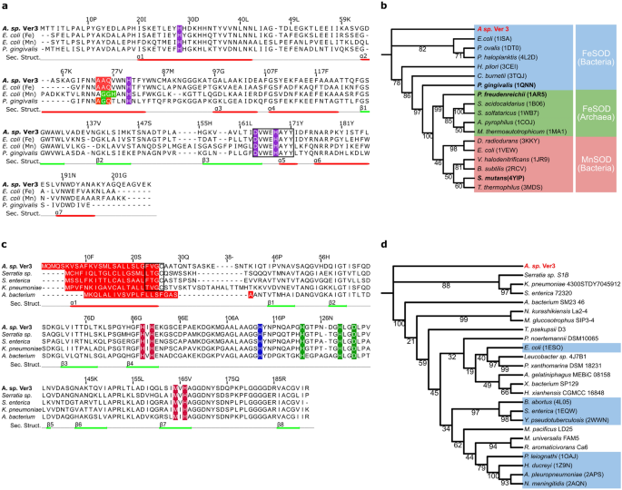
The putative proteins encoded by the sodB and sodC genes from Acinetobacter sp. Ver3 (AV3SodB and AV3SodC, EZQ10255.1 and EZQ12222.1, respectively) have been used as queries in BLASTp searches. The closest match of AV3SodB within the PDB was the FeSOD from Pseudomonas putida (63% id, PDB code 3SDP). Determine 2a shows a a number of sequence alignment of AV3SodB and three structurally characterised associated enzymes: the FeSOD (PDB code 1ISA) and MnSOD (PDB code 1VEW) from E. coli (60% and 44% amino acid id, respectively) and the cambialistic SOD (PDB code 1QNN) from P. gingivalis (50% id). It exhibits that AV3SodB harbours motifs which might be attribute of FeSODs, such because the motifs AAQ and DVEWHAYY concerned in catalysis22. A phylogenetic tree constructed for AV3SodB and numerous FeSODs, MnSODs in addition to cambialistic Fe/MnSODs (right here collectively known as Fe-MnSODs, with greater than 30% sequence id in comparison with AV3SodB and 80% question protection) of recognized construction consisted of three completely different clades, with archaeal and bacterial FeSODs grouped in two completely different clades whereas bacterial MnSODs constituted a 3rd one (Fig. 2b). As anticipated, AV3SodB clustered with bacterial FeSODs. Alternatively, the closest match of AV3SodC within the PDB was the CuZnSOD from Haemphylus ducrey (53% id, PDB code 1Z9N). The sequence alignment in Fig. 2c highlights the conservation of metallic binding residues in AV3SodC and consultant CuZnSODs retrieved from the NCBI database.
Sequence alignment and phylogenetic evaluation of SOD enzymes. (a) Sequence alignment of FeSOD enzymes from chosen species. Amino acids highlighted in purple correspond to conserved metallic ligands. The motifs AAQ and GGH (in MnSODs, the E. coli enzyme is proven for comparability) are proven in purple and inexperienced, respectively; the motif DVWEHAYYID comprising metallic binding residues is boxed. Secondary construction components noticed within the crystal construction of AV3SodB are depicted as purple and inexperienced strains beneath the sequence alignment. A number of sequence alignments have been carried out utilizing MAFFT model 7.475 (https://mafft.cbrc.jp/alignment/software/) with default parameters. (b) Phylogenetic tree of Fe-MnSODs of recognized construction. A clade of MnSODs (purple) and two of FeSODs (blue and inexperienced) are distinguished. Cambialistics Fe/MnSODs are in daring. (c) Sequence alignment of CuZnSOD enzymes from chosen species. Conserved metallic ligands are proven in color: copper ligands in vermillion, zinc ligands in inexperienced and a histidine residue that coordinates each metallic ions in blue. Predicted sign peptides are signalled in purple and lipobox sequences are boxed. Secondary construction components predicted by Jalview60 are depicted as purple and inexperienced strains beneath the sequence alignment. (d) Phylogenetic tree constructed for CuZnSOD sequences retrieved from the NCBI or the PDB (mild blue packing containers). Word that the sign peptide was faraway from every sequence previous to alignment. Timber have been constructed primarily based on the amino acid sequence alignments of the Acinetobacter sp. Ver3 SOD enzymes through the use of the Most probability technique (with a WAG + I + G4 substitution mannequin). The bioinformatic software program IQ-TREE multicore model 1.6.1161 was employed to generate each timber and their reliability was examined by bootstrapping with 10,000 repetitions. Outcomes have been visualized utilizing the net toll iTOL V5.7 (https://itol.embl.de/).
Since FeSODs and CuZnSODs can current completely different subcellular places10, we determined to analyse whether or not AV3SodB and/or AV3SodC introduced sign sequences. No sign peptide was discovered for AV3SodB utilizing the Sign P 5.0 algorithm 23, suggesting that this enzyme is probably going cytosolic. In distinction, AV3SodC displayed a 26-amino acid hydrophobic N-terminal sequence that’s typical of sign peptides for protein export24 (Fig. 2c). A phylogenetic tree calculated from the anticipated mature types of AV3SodC and a wide range of CuZnSODs accessible on the PDB and the NCBI databases (a minimum of 57% sequence id and 70% question protection with AV3SodC) identified that AV3SodC clusters with CuZnSODs from the gammaproteobacteria Serratia sp. S1B, Salmonella enterica and Klebsiella pneumoniae (Fig. 2d). Notably, enzymes belonging to this clade constantly show a putative lipoprotein sign peptide with a predicted lipobox cysteine residue24,25 (Fig. 2c).
AV3SodB and AV3SodC-p are energetic enzymes
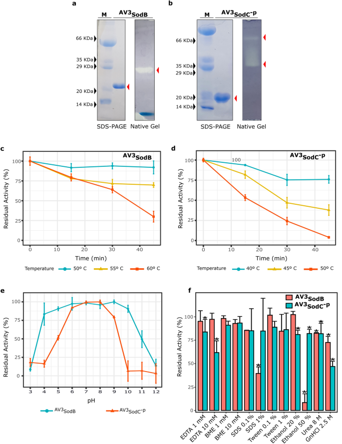
To advance on their characterization, we determined to provide AV3SodB and AV3SodC by recombinant means. AV3SodB was produced in a SOD-deficient E. coli pressure and purified from the soluble cell fraction. The exercise of AV3SodB was assessed by in situ staining after nondenaturing PAGE, as beforehand described26. The obtained outcomes (Fig. 3a) confirmed that AV3SodB is an energetic enzyme. Apart from, the electrophoretic mobility of the energetic AV3SodB species matches that of the one SOD exercise detected in soluble extracts of Acinetobacter sp. Ver3 (not proven).
Biochemical characterization of recombinant Acinetobacter sp. Ver3 SODs. AV3SodB (a) and AV3SodC-P (b) have been analysed by SDS-PAGE and Coomassie Blue staining (left panels). SOD exercise was revealed in situ in nondenaturing polyacrylamide gels (proper panels). M: molecular weight marker. The SDS-PAGE and the polyacrylamide gels have been cropped to create the determine and the full-length gels are introduced in Supplementary Determine S5. (c) Thermostability of AV3SodB and (d) AV3SodC-P. In every case, the residual SOD exercise was measured after completely different warmth remedies. (e) pH tolerance of AV3SodB (blue line) and AV3SodC-P (purple line). In every case, the residual SOD exercise was measured at pH 7.8 after incubating the enzyme 1 h at 25ºC in buffers with completely different pH values. (f) Impact of numerous chemical brokers on AV3SodB and AV3SodC-P exercise. In every case, the residual SOD exercise was measured after incubating the enzyme 1 h at 25º C with the compound. In all experiments, SOD exercise was decided spectrophotometrically by inhibition of the xanthine/xanthine oxidase-induced discount of cytochrome c, at 25º C; the exercise of the untreated enzyme was outlined as 100%. The reported values correspond to the imply of 4 measurements in three replicates of the experiment; bars point out normal deviations.
We tried to provide full-length AV3SodC equally to AV3SodB, nonetheless these efforts repeatedly failed since a number of proteolytic fragments of the protein have been present in SDS-PAGE analyses (not proven), suggesting a doable processing of the N-terminal peptide. Subsequently, we assayed the manufacturing of a AV3SodC truncated model missing the 26-amino acid hydrophobic N-terminal sequence (AV3SodC-p). AV3SodC-p was efficiently produced in E. coli and purified from the soluble cell fraction. AV3SodC-p exhibited two energetic species in non-denaturing gels, most probably corresponding to 2 completely different oligomeric states of the enzyme10 (Fig. 3b).
The particular actions of each Acinetobacter sp. Ver3 SOD enzymes have been decided by the xanthine oxidase technique (as described within the Strategies part), leading to values of 6,600 ± 200 U/mg and 1,800 ± 200 U/mg for AV3SodB and AV3SodC-p, respectively.
Biochemical characterization of AV3SodB and AV3SodC-p
To judge the potential purposes of Acinetobacter sp. Ver3 SODs in business, we examined the results of numerous bodily and chemical effectors on enzyme exercise. The thermostability of recombinant AV3SodB and AV3SodC-p was investigated by pre-incubating the enzymes at completely different temperatures adopted by measurements of the residual exercise (Fig. 3c,d). Notably, the exercise of AV3SodB was just about unaffected after a forty five min warmth remedy at 50 °C. The enzyme even retained about 70% exercise after 45 min at 55 °C and 65% exercise after 30 min at 60 °C. Alternatively, AV3SodC-p conserved solely 80% and 40% of its exercise after incubation at 45 ºC for 15 min or 45 min, respectively. Remedies at larger temperature resulted in a extra pronounced lack of exercise. These outcomes indicated that AV3SodB is extra thermostable than AV3SodC-p.
The residual exercise of AV3SodB and AV3SodC-p was additionally measured after incubation in buffers with pH starting from 3.0 to 12.0. AV3SodB displayed a exceptional stability within the pH vary 4.0–10.0, the place it retained greater than 75% of its exercise (Fig. 3e). AV3SodC-p, then again, conserved greater than 75% of exercise in a narrower vary, between pH values 6.0 and 9.0, declaring that it’s extra delicate to pH modifications than AV3SodB.
EDTA and β-mercaptoethanol (BME) (1 or 10 mM) have been assayed as inhibitors of AV3SodB and AV3SodC-p (Fig. 3f). AV3SodB conserved greater than 90% of its exercise within the presence of each compounds. Nevertheless, whereas AV3SodC-p displayed excessive tolerance to BME, EDTA had an inhibitory impact, resulting in a 40% discount of enzyme exercise when used at 10 mM. SDS and Tween 20 (0.1% v/v or 1% v/v) have been used to analyze the affect of detergents on enzyme exercise (Fig. 3f). SDS severely impaired AV3SodB exercise when used at 1% v/v not like the case of AV3SodC-p the place this agent didn’t intervene with AV3SodC-p exercise. All different results have been negligible. The denaturants urea and guanidine hydrochloride have been additionally assessed, at last concentrations of 8 M and a pair of.5 M, respectively. AV3SodC-p misplaced roughly half of its exercise when handled with guanidine hydrochloride. In all different circumstances enzyme actions remained larger than 70%. To check the soundness of Acinetobacter sp. Ver3 SODs in an natural solvent, the enzymes have been incubated in response buffer supplemented with 20% v/v or 50% v/v ethanol. AV3SodC-p retained nearly 80% of its exercise in each circumstances whereas AV3SodB misplaced almost all its exercise in 50% v/v ethanol.
Total, these outcomes point out the next thermal stability and a broader pH vary for AV3SodB. Conversely, remedy with inhibitors confirmed blended results on enzyme exercise, with AV3SodB being altered primarily by SDS and ethanol, whereas AV3SodC-p was affected by EDTA and guanidine hydrochloride.
Structural characterization of AV3SodB
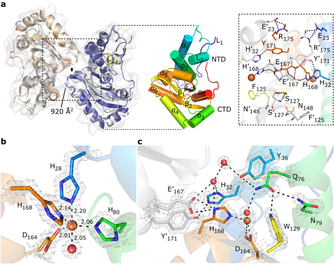
With a purpose to discover the structural properties of AV3SodB, we solved the crystal construction of the enzyme to 1.34 Å decision (Desk 1). AV3SodB crystallized in area group C2221, with one protein molecule per uneven unit. The ultimate atomic mannequin includes AV3SodB residues 1–208. Two neighbouring AV3SodB monomers associated by crystallographic operations kind a protein dimer with cyclic symmetry C2, resembling the quaternary construction of E. coli FeSOD (PDB code 1ISA) (Fig. 4a). The Root-mean-square deviation (RMSD) between the AV3SodB monomer and the chain A within the E. coli FeSOD construction is 0.9 Å for 191 aligned residues. The AV3SodB monomer adopts the two-domain fold conserved amongst FeSODs and MnSODs10, with a helical N-terminal area and a C-terminal area composed of three β-sheets surrounded by α-helices (Figs. 2a and 4a). Per a dimeric useful state, the loop L2 (residues 43–68) that connects the 2 most important helices within the N-terminal area of AV3SodB, essentially the most variable within the main sequence throughout species, not like tetrameric enzymes27,28 isn’t prolonged and establishes hydrophobic interactions with the N-terminal loop L1 (residues 1–19) and the C-terminal area (Figs. 2a and 4a). The interfacial space between AV3SodB monomers is ca. 920 Å2 and the residues concerned in hydrogen bonds or salt bridges between subunits are Glu23, His32, Phe125, Ser127, Asn148, Glu167, His168, Tyr171 and Arg175, all conserved in E. coli FeSOD (Figs. 2a and 4a).
Crystal construction of AV3SodB. (a) Though there’s a single molecule of AV3SodB per uneven unit, two contiguous molecules (in beige and blue, the interface space between them is knowledgeable in Å2) associated by crystallographic symmetry (oval image) give rise to a dimer of the protein which presents a quaternary construction much like that noticed for the E. coli FeSOD (PDB code 1ISA, in Grey, superimposed to AV3SodB) (on the left). Proteins are proven in ribbon illustration. The floor of AV3SodB is depicted in grey. On the proper, a monomer of AV3SodB is proven in rainbow colors, with helices represented as cylinders. Secondary construction components conserved in FeSODs are indicated (see additionally Fig. 2a). NTD, N-terminal area; CTD, C-terminal area. An inset of the interface between AV3SodB molecules in a protein dimer is proven. Residues concerned in intermolecular hydrogen bonds or salt bridges are depicted in stick illustration. Iron ions are proven on this and subsequent panels as orange spheres. (b) Lively web site of AV3SodB. Steel ligands are represented as sticks. Interatomic distances are knowledgeable (in Å). One of many axial ligands is a water molecule (purple sphere). (c) A conserved community of hydrogen bonds involving energetic web site residues. Interatomic interactions are depicted as dashed strains. The mesh in panels (b) and (c) corresponds to the crystallographic 2mFo–DC electron density map contoured to 2.0 σ.
The energetic web site of AV3SodB, positioned near the dimer interface, consists of conserved residues amongst FeSODs (Figs. 2a and 4b). mFo-DFc electron density maps clearly revealed the presence of a metallic ion certain on the energetic web site, which, primarily based on our bioinformatic evaluation, was modelled as an iron ion. The metallic ion is coordinated in a distorted trigonal bipyramidal geometry with the facet chain of His28 and a solvent molecule as axial ligands and the facet chains of His80, Asp164 and His168 as equatorial ligands (Fig. 4b). Residues His28 and His80 are positioned in helices ɑ1 and ɑ2, respectively, of the N-terminal area of AV3SodB, whereas Asp164 and His168 are positioned within the strand β3 of the C-terminal area and the loop that follows it (Figs. 2a and 4b). AV3SodB shares with E. coli FeSOD six out of seven residues at or close to the energetic web site which have been recognized as specificity signature positions for metallic ion use10.
The OH–/H2O ligand hydrogen bonds Asp164 and the energetic web site Gln76 (Fig. 4c). This OH–/H2O, believed to be OH– in oxidized FeSODs and H2O within the lowered enzymes10, is related to bulk water by way of a conserved hydrogen bonding community that begins with Gln76 and continues with residues Tyr36 and His32. The facet chain of Gln76 is stabilized by hydrogen bonding interactions with residues Tyr36, Asn79 and Trp129. Extra conserved hydrogen bonds hyperlink the energetic web site to the interface between AV3SodB monomers, together with a hydrogen bond between His32 of 1 monomer and Tyr171 of the opposite, and a hydrogen bond between His168 of 1 monomer and Glu167 of the opposite.
Apparently, AV3SodB crystallized within the presence of flavin mononucleotide (FMN), added as an additive in selfmade crystallization circumstances. The isoalloxazine ring, clearly evident in mFo-DFc maps, is buried in a deep pocket shaped by helix ɑ2 within the N-terminal area, loops L1 and L2 and essentially the most C-terminal section of the protein (Fig. S1). Notably, much like the FeSOD from H. pylori29, the C-terminus of AV3SodB is longer than for different associated enzymes (Fig. 2a). The isoaloxazine ring of the FMN molecule is positioned at 12.4 Å of the metallic ion and it’s doable to estimate an electron switch path30 between them by means of the His33 and His80 residues (Fig. S1). Digital absorption measurements after incubating AV3SodB with FMN and eradicating the surplus cofactor by measurement exclusion chromatography didn’t, nonetheless, give clear proof of a binding occasion in resolution (not proven). Moreover, it isn’t evident what organic position the binding of FMN to this enzyme might have. To our data, the binding of FMN to a superoxide dismutase has not been beforehand reported. Though the binding of FMN to AV3SodB within the crystal may be very intriguing, this might merely be an artifact and any additional exploration of this remark would require particular experiments.
AV3SodB is a cytosolic enzyme whereas AV3SodC is directed to the bacterial periplasm and loaded into OMVs
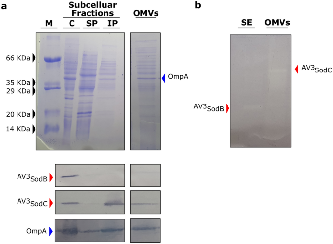
To find out the subcellular localization of AV3SodB and AV3SodC, we obtained polyclonal antibodies in opposition to the recombinant proteins and used them to carry out Western Blot analyses of Acinetobacter sp. Ver3 cell fractions. 4 completely different fractions have been ready: cytosol, soluble periplasmic fraction, insoluble periplasmic fraction and outer membrane vesicles (OMVs). AV3SodB was detected within the bacterial cytosol whereas AV3SodC was moreover discovered within the insoluble periplasmic fraction (Fig. 5a). Per the subcellular localization of AV3SodC, its N-terminal sequence includes a (−3)[LVI](−2)[Xaa](−1)[Yaa](+1)C motif (Fig. 2c) that could be a hallmark of bacterial lipoprotein attachment websites24,25, suggesting that AV3SodC could possibly be related to the membrane.
AV3SodB and AV3SodC subcellular localization. (A) Coomassie stained SDS-PAGE (higher panel) and Western Blot assay (decrease panel) of cytosolic (C), soluble and insoluble periplasmic (SP and IP, respectively) fractions (7 µg of complete proteins), and OMVs (15 µL from a 500X tradition focus) obtained from Acinetobacter sp. Ver3. Particular antibodies raised in opposition to AV3SodB and AV3SodC (purple arrows) have been used. The detection of OmpA (blue arrow) with particular antibodies in opposition to the A. baumannii protein was used as a management; OmpA has been proven to be loaded into OMVs31. (B) In-gel evaluation of SOD exercise current in a soluble extract (SE) (1.5 µg of complete proteins) and OMVs (15 µL from a 500X tradition focus) of Acinetobacter sp. Ver3. M: molecular weight marker. The specificity of the antibodies was examined exhibiting that there isn’t any cross-reactivity (Fig. S4). The SDS-PAGEs and the blots have been cropped to create the determine. Full-length gels/blots are introduced in Supplementary Determine S5.
For the reason that CuZnSOD from A. baumannii ATCC 17978 has been detected in OMVs32, we investigated whether or not this was additionally the case for AV3SodC. Certainly, AV3SodC was present in OMVs obtained from Acinetobacter sp. Ver3 cultures (Fig. 5a). Notably, AV3SodC is energetic when positioned in OMVs (Fig. 5b).
Differential expression of sodB and sodC in response to pro-oxidant challenges
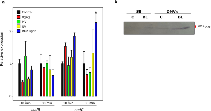
With a purpose to additional discover the physiological roles of AV3SodB and AV3SodC, we studied the modifications in sodB and sodC expression in Acinetobacter sp. Ver3 cells subjected to pro-oxidant challenges. qRT-PCR analyses have been carried out from complete RNA samples obtained 10 and 30 min after the publicity of cells to MV, H2O2, UV radiation and blue mild. The latter remedy was carried out as a result of blue mild acts as a pro-oxidant agent in micro organism by producing ROSs33. Moreover, blue mild induces catalase exercise in Acinetobacter baumannii34, and thus might play an essential position within the defence in opposition to oxidative stress.
The expression of sodC elevated about twofold after a 30 min blue mild problem, however no vital modifications have been noticed beneath the remainder of the pro-oxidant circumstances (Fig. 6a). In distinction, sodB transcript ranges remained unaltered beneath the circumstances examined. To find out whether or not these outcomes confirmed a correlation on the protein stage, exponentially rising cell cultures have been uncovered to blue mild and protein extracts have been then obtained. Soluble fractions in addition to OMVs have been analysed by SDS-PAGE adopted by anti-AV3SodC immunostaining (Fig. 6b). A internet accumulation of AV3SodC was noticed in OMVs after the blue mild remedy.
sodB and sodC response to pro-oxidant challenges. (a) Relative ranges of sodB and sodC transcription in untreated Acinetobacter sp. Ver3 cells (black bars) or after publicity to 1 mM H2O2 (purple bars), 0.5 mM Methyl Viologen (MV) (inexperienced bars), 900 J.m-2 ultraviolet (UV) (yellow bars) or blue mild (blue bars). The imply for the housekeeping genes recA and rpoB was used as a normalizer. Asterisks point out vital variations amongst management and handled samples, as decided by evaluation of variance (ANOVA) and Tukey’s a number of comparability take a look at. In every case, the typical worth and the usual deviation of 4 organic replicates are proven. (b) Anti-AV3SodC immunoblot of a soluble extract (SE, 7 µg of complete proteins) and OMVs (15 µL from a 500X tradition focus) of Acinetobacter sp. Ver3 grown over 26 h within the presence of blue mild (BL, 5 μmol.m-2.s-1). A management (C) tradition was additionally included. The total-length blot is introduced in Supplementary Determine S5.
Prevalence of SOD-encoding genes within the Acinetobacter genus
With a purpose to discover the range of SOD-encoding genes within the Acinetobacter genus, we assembled an area database with 314 publicly accessible full genomes, comprising 53 Acinetobacter outlined species in addition to 21 strains similar to unassigned species (Desk S1). BLASTp searches have been then carried out utilizing the sequences of the enzymes FeSOD (ABO12765.2)19 and CuZnSOD (ABO13540.2) from A. baumannii ATCC 17978 as queries. We discovered 677 proteins (Desk S2), and by inspecting the SOD-encoding genes within the chosen Acinetobacter strains, we distinguished three genotypes (Desk S1):
-
1.
Genotype 1 (1 FeSOD + 1 CuZnSOD; 83.4% of the strains). This group accommodates representatives of 28 species, however it’s remarkably enriched in these which have been related to nosocomial infections, comparable to A. baumannii, A. pittii and A. nosocomialis. It additionally consists of 17 unassigned strains, comparable to Acinetobacter sp. Ver3. CuZnSOD enzymes on this group include a predicted lipoprotein sign peptide and will thus be translocated to the periplasm or the extracellular area equally to AV3SodC. We additionally embody on this group three A. baumannii strains (921, 3207, 7835), bearing two FeSOD encoding genes, which have been denominated sodB1 and sodB2.
-
2.
Genotype 2 (1 FeSOD + 1 MnSOD + 1 CuZnSOD; 10.2% of the strains). This class encompasses a complete of 18 Acinetobacter species and three unassigned strains. MnSODs in 27 out of 32 strains of this group are predicted to be cytosolic enzymes, apart from one A. beijerinckii and three A. junii strains, which encode MnSOD variants with a predicted sign peptide and not using a lipidation motif (Fig. S2). Apparently, A. bereziniae XH901 encodes MnSODs of the 2 varieties, comprising a complete of 4 SOD encoding-genes within the genome. Much like genotype 1, CuZnSOD enzymes on this group include a predicted lipoprotein sign peptide.
-
3.
Genotype 3 (1 FeSOD + 1 or 2 MnSOD; 6.1% of the strains). This group accommodates 7 species comprising a complete of 18 strains, with A. haemolyticus essentially the most prevalent species (11 strains). One pressure similar to an Acinetobacter unassigned species can be included on this group. Remarkably, all strains encode a MnSOD enzyme containing a putative sign peptide and not using a lipidation motif (Fig. S2), which could possibly be directed to the periplasmic area to play a CuZnSOD-like position. Twelve strains moreover bear a putative cytosolic MnSOD.
An uncategorized case is that of the species A. apis, which encodes 1 CuZnSOD and 1 MnSOD (Desk S1), however lacks a FeSOD-encoding gene. The MnSOD is predicted to be cytosolic (Fig. S2).


