Plackett–Burman media screening for chemically outlined medium growth
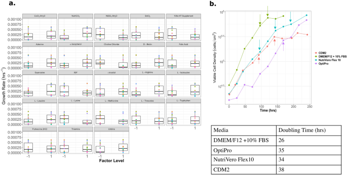
4 Plackett–Burman styled experiments have been carried out to quickly display media parts to help suspension progress in Vero cells ranging from a DMEM/F12 base. The main carbon supply was glucose, which was elevated from 17.5 to 25 mM, and the l-alanyl-l-glutamine focus was elevated to 4 mM. In complete, 62 compounds have been examined over 96 formulations within the 4 completely different Plackett–Burman experiments (see Supplementary Desk S1). From this, a chemically outlined adherent and suspension medium formulations (CDM) have been made for Vero cells. The medium formulations have been improved iteratively with every subsequent Plackett–Burman experiment, and the bottom formulation for the following experiment was designed from the earlier set of outcomes. An instance of the outcomes from a Plackett–Burman experiment designed for suspension media is illustrated in Fig. 1a, the place the impact of every element on progress price is proven. Via this experiment, the impact of purines and pyrimidines (adenine, guanosine, uridine, and HT complement) may be seen having total a optimistic impact on progress price, aside from the HT complement which was supplemented at too excessive of a focus for the very best issue degree. One other class of compounds that improved the expansion price was metals. Along with metals comparable to calcium, magnesium, iron, copper and zinc, varied hint metals have been added to the medium formulation. Particularly cobalt, maganese, molybdenum, silicate, selenite, nickel, tin, and vanadium, that are usually excluded from basal media, however may be present in serum, have been important for cell viability and progress12. Hint metals are vital cofactors in varied enzymes, and these metals may be present in deionized water, however not in extremely pure water. One of many drawbacks of including metals to protein-free media, is that metals are pro-oxidants and might result in the oxidation of media if antioxidants or metallic chelators will not be current. Antioxidants comparable to α-tocopherol (Vitamin E), ascorbic acid (Vitamin C), glutathione, citric acid, and pyruvate have been added to the medium. Of those compounds, all of them have been useful at excessive concentrations, aside from glutathione, which had a detrimental impact on cell progress at concentrations larger than 3.3 µM (Supplementary Fig. S3). All of the experiments and the focus of every issue degree may be discovered within the Supplementary Figs. S1–S4.
(a) Field plots that present the impact of every media element for a Plackett–Burman experiment with 23 elements in 24 runs. This experiment used a basal media with low calcium and magnesium to encourage suspension progress. The expansion price was use because the dependent variable to match the affect of every media element. (b) The adherent chemically outlined media (CDM) formulation was in comparison with business media. The arrows point out when media was exchanged. CDM2, CDM3, and CDM4 have been formulated primarily based on the outcomes from Plackett–Burman experiments 4, 6 and seven, respectively.
Development elements, polyamines, nutritional vitamins and steroids have been investigated to extend the expansion price. Surprisingly, not all progress elements had a optimistic impact on the expansion price of Vero cells, insulin-like progress issue (IGF) had no poistive impact, whereas recombinant epidermal progress issue (rEGF) was required for progress (Fig. 1 and Supplementary Fig. S1). Newer media formulations developed for Vero cells have used rEGF13,14 fairly than fibroblast progress issue (FGF). Cinatl et al., discovered that Vero cells may develop properly on polyvinyl formal flasks in a protein free media that contained progesterone15. It’s possible that rEGF could possibly be changed by a mixture of steroids to create a protein-free medium for Vero cells.
The adherent chemically outlined medium contained 10 ng/mL of rEGF as the one protein current within the remaining formulation and achieved a doubling time of 32 h, in comparison with OptiPRO SFM (35 h) and NutriVero Flex10 (34 h) (Fig. 1b). Epidermal progress issue (EGF) is often added to chemically outlined cell tradition media as a result of the EGF receptor (EGFR, also referred to as ErbB1) is expressed on virtually all cell sorts. Plant hydrolysates have been recognized to imitate progress factor-like results for a lot of completely different cell sorts at low price and is animal component-free (ACF), which makes them a great different to supplementing with particular person progress elements16,17,18. OptiPRO SFM was chosen as a comparability as a result of it’s generally utilized in trade as a medium to provide vaccines utilizing Vero cells. This medium is animal component-free but it surely does comprise a really low protein focus of plant hydrolysates (≤ 10 μg/mL)19. NutriVero Flex10 is one other Vero cell medium that’s commercially obtainable, and in contrast to OptiPro, it’s chemically outlined, in addition to ACF, making it a greater comparability to the medium developed on this paper.
Total, not one of the ACF media formulations (NutriVero Flex 10, OptiPRO SFM, or CDM2) grew as quick as serum-containing DMEM/F12 with 10% FBS. Due to this fact, one may infer that there are nonetheless some parts lacking from ACF media comparable to cell signaling molecules which can be in low concentrations, or progress elements which can be lacking from serum-free media. A research carried out by Desai et al., tracked the expansion elements that have been produced by Vero cells20,21. They discovered that Vero cells excreted platelet derived progress issue (PDGF), interleukin 6 (IL-6) and leukemia inhibitory issue (LIF), however have been unable to detect EGF or lively TGF-β. Apparently, Guo et al., have been solely capable of finding EGFR on Vero cells after they carried out a proteomic evaluation of the membrane proteins22. This may increasingly have been because of the restricted annotation of the Vero genome and floor proteins, or that the EGFR in Vero cells can work together with many alternative ligands. Nonetheless, rEGF did stimulate proliferation of Vero cells adherently within the chemically outlined medium, however the literature means that different progress elements could possibly be used as well as, or to exchange rEGF.
Suspension medium growth
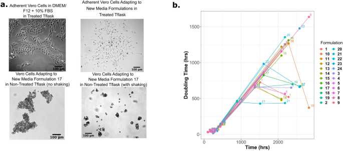
Cells have been slowly tailored from DMEM/F12 + 10% FBS medium in adherent tradition to the brand new medium formulations, and because the quantity of calcium and magnesium decreased, the cell progress price was additionally noticed to lower dramatically. On the lowest focus vary of 0.1 mM calcium, Vero cells have been nonetheless in a position to adhere to the tissue-culture handled Tflasks; subsequently, cells have been transferred to non-treated Tflasks. Tissue tradition handled Tflasks are generally coated with polylysine, however Tflasks will also be coated with collagen, laminin or Matrigel to encourage cell attachment23,24. Via subsequent passaging in non-treated Tflasks, Vero cells indifferent and shaped giant aggregates (Fig. 2a). After 90 days of culturing in non-treated Tflasks, the cells have been moved onto a shaker at 40 rpm in an effort to interrupt up the cell aggregates. Just one formulation supported viable single cells (Formulation 17).
(a) Brightfield pictures of Vero cells as they adapt to low-calcium and magnesium media over the course of 180 days. Vero cells grown in DMEM/F12 + 10% FBS is used as a reference for regular morphology, in comparison with the cells rising within the chemically outlined media (prime proper picture). Vero cells have been unable to stick to non-treated T-flasks within the chemically outline media, and with the addition of shaking (40 rpm, backside proper picture) single cells and small aggregates have been achieved. (b) The doubling time of Vero cells elevated dramatically as they have been tailored to the low-calcium and magnesium media over the course of 118 days. The shortest doubling time on the finish of the experiment was roughly 20 days (500 h).
Sodium bicarbonate (NaHCO3) had a big impact on the doubling time, which may be seen in Fig. 2b. There are two main teams in Fig. 2b, one group has a big doubling time (> 1000 h), and one with a shorter doubling time (~ 500 h). The sooner rising group contained 2.2 g/L of sodium bicarbonate, whereas the slower group had 1.2 g/L. For five% CO2 incubators, the focus of NaHCO3 needs to be between 1.2 and a pair of.2 g/L, and three.7 g/L for 10% CO2 incubators25. For our medium formulation, 2.2 g/L of sodium bicarbonate offered additional buffering. Even with the enhancements seen from including extra NaHCO3, the doubling time of those cells was very prolonged (doubling time of 20 days), subsequently additional medium formulations centered on lowering the doubling time.
One of many main variations between adherent media and suspension media is the focus of calcium and magnesium. These two ions are vital for cell adhesion proteins comparable to cadherins, which require a sure focus (> 55 μM) of calcium to take care of the proper inflexible conformation to stay lively26. Calcium can also be concerned in lots of mobile processes apart from cell adhesion27, comparable to cell signaling28, enzyme exercise, and apoptosis29. Wholesome cells preserve a big focus gradient between the cytosol (0.1 μM) and extracellular house (1–2 mM)27. Magnesium is the second (after potassium) most ample cation contained in the cell and ranges from 17 to twenty mM in most mammalian cells30. A lot of the magnesium is sure to varied mobile buildings, and solely about 0.8–1.2 mM is free Mg2+30. It’s important for essentially the most fundamental capabilities within the cell, for instance, for ATP to be biologically lively. Low Mg2+ focus has additionally been linked to accelerated differentiation of bone-marrow-derived mesenchymal stem cells (MSCs) into osteoclasts by means of elevated reactive oxygen species (ROS) technology31. Whereas for adipose-derived MSCs the ten × discount (1 mM Mg, versus 0.1 mM Mg) of Mg in reprogramming medium triggered the cells to have elevated expression of genes related to MSCs (gata-4, nkx-2.5, hgf, kdr, nerog, nanog)32. It’s almost certainly that the discount of those ions within the suspension medium (Formulation 17) had detrimental results on cell signaling, DNA replication, RNA transcription, and enzymatic exercise. Doubtlessly, the low magnesium ranges could possibly be inflicting transcriptional variations within the suspension cells which can be inflicting them to distinguish. Subsequently, Formulation 17 was modified to have the identical focus of calcium and magnesium ions as DMEM/F12 for comparability functions, and was referred to as CDM2. Formulation 17 was thus renamed ‘CDM2 low calcium/low magnesium’ henceforth.
Development with different cell strains
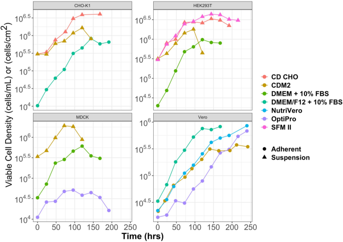
To analyze if CDM2 may help progress of different cell strains, MDCK, CHO-K1 and HEK293T cells have been sequentially tailored to CDM2 from serum-containing basal medium utilizing static Tflasks. HEK293T and MDCK cells have been tailored over 2 weeks, whereas CHO-K1 cells took 3.5 weeks to adapt to CDM2. On condition that CDM2 accommodates the identical quantity of calcium and magnesium as basal DMEM/F12, this indicated that cells may nonetheless develop in suspension with greater ranges of the divalent cations. Determine 3 demonstrates how the cells grew in CDM2 in comparison with business media, and serum-containing basal media, together with the utmost cell densities achieved when culturing the cells in suspension. For CHO-K1, HEK293T and MCDK cells grown in CDM2, cells began to lose their viability after roughly 4 days. After day 3, a major colour change within the medium could possibly be seen indicating that the media was turning into acidic. On condition that this medium was developed for adherent Vero cells, the flexibility to develop 3 different cell strains in suspension was a stunning end result that led to the query of what’s completely different about Vero cells that stops them from rising properly in suspension.
The chemically outlined media that was formulated from the sequence of Plackett–Burman experiments was modified to incorporate 1 mM Ca2+ and a pair of mM Mg2+ (referred to as CDM2) to help the next progress price in Vero cells. CHO-K1, HEK293T and MDCK cells have been slowly tailored to CDM2 from serum-containing basal media. All three cell sorts have been in a position to develop in suspension (triangle) after they have been totally tailored to CDM2 and have been cultured in shaker flasks. Vero cells remained adherent for all media formulations. Suspension (stuffed triangle), adherent (open circle).
RNA-seq experiment to mobile expression modifications
To determine the explanations for the lowered progress of the Vero cells in suspension in an outlined medium, a transcriptomic evaluation was carried out to determine expressional modifications. This information helped to determine metabolic processes and transcription elements that would result in a decreased doubling time within the chemically outlined medium. The RNA-seq dataset comprised three teams of cells; (1) Vero cells grown adherently in DMEM/F12 + 10% FBS, (2) Vero cells grown adherently in CDM2 with added calcium and magnesium, or (3) Vero cells grown in suspension in CDM2. Cells grown adherently in DMEM/F12 medium (Management, or ‘Con’) and cells grown adherently in CDM2 with further calcium and magnesium (Adherent, or ‘Adh’) have been sequenced and in comparison with cells grown in suspension in CDM2 (Suspension, or ‘Sus’). A abstract of the alignment statistics is offered in Supplementary Desk S2. Over 96% of the reads for every pattern have been uniquely aligned to the Chlorocebus sabaeus (C. sabaeus) genome. After filtering the recognized genes for an expression of at the least 1 CPM in 4 samples, 11,135 genes have been thought-about as expressed for additional evaluation (Supplementary Desk S3). As a result of restricted details about mobile pathways in C. sabaeus, the Ensembl database was used to determine H. sapiens homologs for the expressed genes. This allowed a extra detailed gene set enrichment evaluation. The entire outcomes of the differentially expressed gene evaluation are in Supplementary Tables S4–S7.
The variability inside the dataset was analyzed with: (1) a principal element evaluation (PCA) of the five hundred most variable genes; and (2) a Pearson correlation of 3122 recognized, expressed housekeeping genes (Supplementary Fig. S5). The Pearson correlation of housekeeping genes managed for outliers with unanticipated modifications in housekeeping gene expression. The PCA confirmed a transparent separation between all three circumstances with Adh samples being grouped between Con and Sus samples as predicted.
Modifications in regulation of proliferation and apoptosis
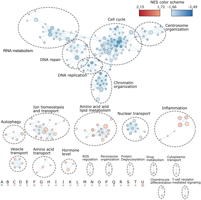
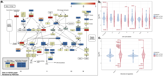
GSEA evaluation was used to determine essentially the most constant expression modifications in gene units. An enrichment map of the numerous down-regulated GO-terms in organic processes with a stringent FDR threshold of 5% may be seen in Fig. 4 (Supplementary Desk S8). Total, 151 gene units have been considerably down-regulated, and 91 have been recognized as considerably up-regulated. Most down-regulated gene units have been associated to cell cycle regulation, mitosis, DNA-replication and DNA group which is according to the remark of the lengthy doubling time of Vero in suspension (Fig. 5a)33,34,35. Suspensions cells had down-regulated genes from every a part of cell cycle development in comparison with adherently grown Vero cells in CDM2 (Fig. 5b). The gene c-myc is down-regulated in suspension Vero cells, and this gene has been related to cell cycle development, together with tumorogenesis36. The overexpression of this gene has been proven to permit quiescent cells to reenter the cell cycle and start to proliferate36. Up-regulated genes have been linked to irritation, fluid shear stress, migration and endothelial boundaries, indicating a stress response throughout cell adaption to suspension. Importantly, gene units associated to regulation of programmed cell loss of life weren’t detected as both down- or up-regulated. Genes units regulating apoptosis weren’t detected as differentially expressed by GSEA, as seen by the expression patterns of optimistic regulatory (GO:0043065) and damaging regulatory (GO:0043066) gene units associated to apoptotic processes (Fig. 5c). Regardless of a wider log fold-change (LFC) vary for pro-apoptotic genes and anti-apoptotic genes, the common LFC for each units of genes is roughly zero for Adh samples, and solely barely optimistic for Sus samples.
Enrichment map for important downregulated GO-terms in organic processes for the comparability of Sus_CDM2 in opposition to Adh_CDM2 (threshold at FDR of 0.05). The scale of the node is proportional to the variety of recognized genes annotated to the GO-term. The thickness of edges represents the extent of overlap between the GO-terms. A: DNA templated transcription termination, B: Cardio respiration, C: Response to elevated oxygen ranges, D: Protein peptidyl prolyl isomerization, E: Cofactor transport, F: optimistic regulation of inflammatory response, G: Mobile response to fluid shear stress, H: Regulation of ubiquitin protein ligase exercise, I: Institution of endothelial barrier, J: Hyperosmotic response, Ok: Regulation of protein oligomerization, L: Excitatory synapse meeting, M: Regulation of calcineurin mediated signaling, N: Modified amino acid transport, O: Cell migration concerned in sprouting angiogenesis, P: Low density lipoprotein receptor particle metabolic course of, Q: Endosome group, R: Pyrimidine nucleotide triphosphate biosynthetic course of, S: Water soluble vitamin metabolic course of, T: Collagen catabolic course of, U: Drug metabolic course of. NES: Normalized enrichment rating; The entire gene set record with group affiliation is proven in Supplementary Desk S8.
Expression regulation of cell cycle associated genes. (a) KEGG cell cycle displaying differentially regulated genes for the comparability of Sus_CDM2 in opposition to Adh_CDM2 rendered with Pathview. The colour code determines the path of considerably differentially regulated genes (FDR < 0.001) based on the legend on the prime of the determine. A white background of a textual content field implies that the gene was not recognized as expressed. (b) LFC for the highest 500 cell cycle associated genes from Cyclebase 3.0 grouped to their peak expression part. Non-periodic: no recognized expressional modifications through the cell cycle, G1: Hole part 1 of the cell cycle, G1/S: Hole 1 to synthesis transition part, S: synthesis part, G2: Hole part 2, G2/M: Gap2 to mitosis transition part, M: mitosis part. (c) LFC for genes linked to optimistic (optimistic) or damaging (damaging) regulation of apoptosis based on gene ontology.
The very best LFC for anti-apoptotic genes lrp2, pkhd1 and tmigd1 have been proven to particularly shield renal cells from apoptosis37,38,39. Then again, for the highest up-regulated pro-apoptotic genes, tnfsf10 has been confirmed to set off apoptosis in renal cells. The expression of apoptogenic genes like bax, bak1, or dangerous have been unaffected, whereas, anti-apoptic genes are both unchanged like mcl1 or bcl2l2 or enhance like bcl240. Total, the sample doesn’t appear conclusive for the Sus samples to point a change within the price of apoptosis because of the widespread expression modifications of each pro- and anti-apoptosis regulating gene units.
Mitochondrial fatty acid metabolism
The fatty acid (FA) beta oxidation pathway is the principle up-regulated gene set within the GSEA amino acid and lipid metabolism cluster (Fig. 4). The product of this pathway, acetyl-CoA, is additional transformed within the Krebs cycle for power manufacturing. The expression modifications of FA beta oxidation in Sus samples are offered in Supplementary Fig. S6. The heatmap in Fig. S6b illustrates the upregulation of 29 beta oxidation related genes. The schematic diagram in Fig. S6a illustrates that for every step in fatty acid beta oxidation at the least one gene is up-regulated. This upregulation of enzymes for every most important step may relate to an elevated power consumption. One key regulator of fatty acid metabolism in renal cells is tgfb141 (encodes for reworking progress factor-β1 protein), which is down-regulated in Sus cells whereas its receptor tgfb1r is up-regulated. The expression of the tgfb1 gene inhibits beta oxidation and promotes dedifferentiation, together with the epithelial-to-mesenchymal transition42. Differentiated tubular epithelial cells use FA beta oxidation as their most important technique to provide power41. Apparently, in non-cancerous cells the addition of remodeling progress issue beta (TGF-β) will trigger the down-regulation of c-myc and cease cell proliferation36, however as soon as the cancerous phenotype is already established, TGF-β is unable to cease them from proliferating. In abstract, this information helps the concept suspension Vero cells have gotten quiescent or senescent cells through the use of fatty acid beta oxidation, together with the big doubling instances and down-regulated cell cycle genes. A attainable technique to beat this senescence could also be to up-regulate key genes comparable to c-myc36, bcl243, or the addition of TGF-β44.
Upregulation of kidney associated genes
An enrichment map for up-regulated GO-terms for organic processes confirmed three giant clusters of upregulated gene units (Fig. 4). These gene units are associated to ion and small natural molecule transport in addition to amino acids and lipid metabolism. To determine if the modifications within the Sus group have been linked to tissue particular sample and tissue profiles for enriched genes have been outlined from the human protein atlas (HPA, obtainable from http://www.proteinatlas.org) RNA-seq information45. The numerous profiles (p-adj < 0.05) of the gene set enrichment evaluation with the tissue profiles may be seen in Fig. 6a, which reveals that kidney and liver enriched genes have been up-regulated. There have been 17 genes related to the liver expression profile and 12 of them contributed to the enrichment rating, whereas the liver profile from HPA accommodates 242 enriched genes. The kidney profile accommodates 59 genes from which 25 have been recognized within the dataset and 22 contributed to the enrichment rating. The 25 genes are additional proven within the mean-difference (MD) plot in Fig. 6b. Aside from slc13a3, npr3, and lhx1, all genes have been considerably up-regulated with an FDR < 0.001. Since Vero cells have been initially derived from a kidney, and the expression profile seems to particularly match the HPA kidney profile, additional evaluation was executed on kidney-related genes.
(a) HPA tissue associated gene set enrichment evaluation of Sus_CDM2 samples in comparison with Adh_CDM2 samples (FDR < 0.05). Tissue profiles have been developed with the bundle TissueEnrich. Solely tissue enriched genes have been chosen for the profiles, and solely profiles with measurement of at the least 5 particular genes have been thought-about for the evaluation. The proportion of the genes associated to that profile and contributing to the enrichment scores are annotated along side the bars. The coloring of the bars depicts the FDR values based on the legend on the correct facet of the determine. (b) MD plot of recognized kidney enriched genes with LFC and log(CPM) values. Inexperienced and purple labels renal tubules or kidney related genes based on Human Protein Atlas database, respectively. (c) Identification of renal tubule section particular expression sample. Section associated gene set enrichment evaluation of Sus_CDM2 samples in comparison with Adh_CDM2 samples (FDR < 0.1).
Based mostly on this up-regulation of renal tubule related genes, expression modifications have been investigated to see if they could be associated to much more particular segments of the renal tubule. To construct profiles for the completely different segments bulk RNA-seq of dissected rat kidneys have been used46. 13 section profiles have been constructed with TissueEnrich of which eight handed the brink of three detected genes within the RNA-seq dataset. The outcomes are summarized in Fig. 6c (FDR threshold of 0.1). The entire outcomes are in Supplementary Desk S9. The enrichment evaluation presents an up-regulation of a number of units referring to completely different components of the renal tubule with proximal tubule section S3, cortical thick ascending limb which is a part of the Loop of Henle in addition to the inside medullar a part of the accumulating duct (IMCD). In distinction, solely the glomerulus associated gene set was detected as downregulated. From the 25 genes that have been recognized from the dataset, twelve of those genes are particular for renal tubule based on HPA (purple factors in Fig. 6b). Collectively, these findings recommend a mobile change in direction of renal tubule like expression patterns through the adaption to suspension circumstances used on this research.
Moreover, the evaluation of transcription issue goal gene units revealed the up-regulation of a number of kidney related transcription elements (Supplementary Desk S10). The three strongest enriched genes units have been foxi1, pax2, and hnf4a. The foxi1 gene is understood to be vital in regulating the expression of vacuolar-type H+-ATPase subunits within the kidney accumulating duct47; whereas, pax2 is a crucial issue for nephron differentiation in kidney growth48. The primary perform of hnf4a is the management over the expression of drug metabolizing enzymes and transporters within the proximal tubule49. The regulatory community of those transcription elements strongly helps the directed modifications in direction of renal tubule cells.
Vero transition to renal tubule-like cells
Membrane transporter
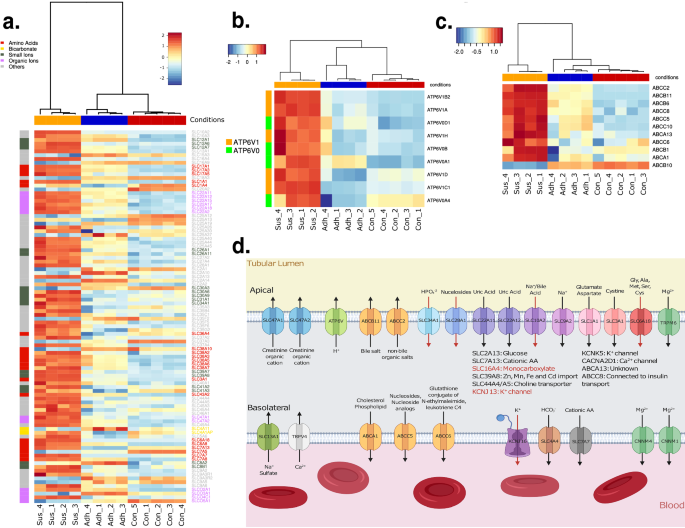
A better investigation of widespread renal tubule capabilities reveals much more similarities with the Sus samples. Membrane transport proteins like these from the solute service (SLC) superfamily are vital in renal tubule cells to move numerous molecules, from ions to lipids or amino acids, to and from the bloodstream or tubular lumen. 246 genes of this superfamily have been recognized as expressed. The heatmap in Fig. 7a reveals the modifications for solute carriers. The elevated expression of many SLC genes point out a change of the Sus cells to elevated absorption and secretion like renal tubule cells.
(a) heatmap of differentially regulated solute service. (b) heatmap of differentially regulated subunits of V-type proton ATP (ATP6V). (c) heatmap of differentially regulated ABC-transporter. (d) Scheme of the considerably up regulated membrane transporter with the goal molecules and the path of transport in a nephron. The transporters whose location are unknown are talked about within the middle of the picture. Pink coloured arrows or gene names point out an LFC > 5 (Created with Biorender.com).
V-ATPases type one other group of transporters whose expression elevated. V-ATPase are proton pumps, which comprise two domains. The heatmap in Fig. 7b emphasizes the robust up-regulation of subunits for these domains in Sus samples, particularly for atpv6v1, which is an ATP hydrolysis area. For atp6v1, 5 of seven subunits are upregulated whereas for atp6v0, two of the eight are up-regulated. The clustering of the samples reveals once more a powerful distinction between Sus samples in opposition to the opposite samples. V-ATPase is vital for proton secretion into the tubular lumen and thus urine acidification, and it’s situated within the apical membrane50,51.
An analogous sample may be seen for drug secreting ATP-binding cassette (ABC) transporters. Ten members, together with abca, abcb, and abcc have been upregulated. Once more, the transporters are strongly upregulated in Sus samples as may be seen in Fig. 7c. The heatmap additionally reveals that the rise in expression occurred progressively from Con to Adh to Sus cells. In renal tubules, ABC transporters assist to secrete medication in addition to a variety of macromolecules from lipids to bile salts or insulin into the tubular lumen52,53,54.
To review the transport path, the renal tubule related carriers are represented in Fig. 7d with substrate and orientation. The diagram reveals a powerful path for transport from many molecules into the cell and from there into the blood stream. The transported goal molecules which can be facilitated from the extremely expressed genes which can be additionally related to transport from the lumen are numerous and embrace bicarbonate, uric acid, magnesium ions and amino acids. Though the Sus samples had no separation between a possible blood stream and a tubular lumen, the RNA-seq information present an up-regulation of direction-specific transmembrane transporters for Mg2+ or amino acids (Fig. 7d). This means that when these Vero cells have been positioned in suspension, the physiological circumstances triggered the cells to revert to a extra kidney-like cell that centered on filtering the medium fairly than proliferating.
Barrier perform between tubular lumen and blood stream
To advertise single cell suspension, CDM2 had low quantities of calcium and magnesium to forestall calcium-dependent cell adhesion molecules from correctly functioning. Many cell adhesion molecules require calcium or magnesium to be practical comparable to cadherins, integrins and selectins. Though, even with decrease calcium and magnesium concentrations, this didn’t stop all cell adhesion since there are different adhesion molecules that don’t require calcium or magnesium like claudins and vascular adhesion molecules (VCAM-1). Claudins are transmembrane proteins that particularly bind cells collectively at tight junctions. Vero cells that have been grown adherently had down-regulated expression of assorted claudins, whereas suspension Vero cells up-regulated many cell adhesion proteins together with claudins (besides cldn15) (Fig. 8). Tight junctions are particularly vital within the kidney tubule to regulate passive reabsorption of explicit compounds between the blood stream and the tubular lumen, which additionally signifies that the suspension Vero cells have been behaving extra like senescent kidney cells55. The upregulation of those proteins, together with different cell adhesion molecules comparable to itgb2, vcam1 and jam1 will be the cause why even with low calcium and magnesium ranges, the cells would nonetheless type aggregates (Fig. 2a).
Heatmap of differentially regulated genes. The x-axis reveals the samples and the y-axis represents the HGNC symbols. The coloring of the heatmap represents the standardized, normalized expression log2CPM values based on the legend on the prime left facet of every heatmap. (a) Warmth map of tight junctions related claudins. (b) Heatmap of differentially regulated drug metabolism genes for part 1 modification. (c) Heatmap of differentially regulated drug metabolism genes for part 2 conjugation.
Conclusion
A chemically outlined media was designed that helps the expansion of assorted mammalian cell strains in suspension. Vero cells have been unable to successfully proliferate in suspension on this medium, and transcriptomic work revealed that these cells have been behaving extra as mature kidney cells. Total, the RNA-seq information proven right here demonstrated that suspension Vero cells grew to become senescent fairly than apoptotic, and that genes related to cell-cycle development have been down-regulated and genes related to fatty acid beta oxidation have been up-regulated. Along with the arrest in progress, suspension Vero cells up-regulated kidney tissue-associated genes. Suspension Vero cells particularly up-regulated genes related to solute membrane transporters and proteins that type tight junctions which is vital for the epithelial barrier and performance of the tubule of the kidney for filtering blood. This work presents insights as to why Vero cells have been troublesome to adapt to suspension prior to now and elucidates some attainable avenues to determine a strong suspension cell line to help vaccine manufacturing utilizing Vero cells.

