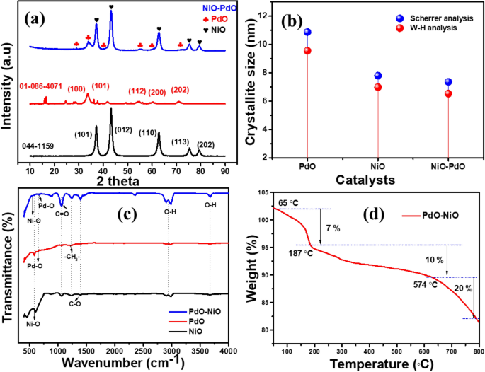
The crystalline nature and part purity of the synthesized NiO, PdO and PdO–NiO nanocomposite have been confirmed by XRD evaluation and proven in Fig. 1a. All three XRD patterns present the extremely crystalline nature, which confirms the purity of the samples. In PdO–NiO blended steel oxide, The extreme diffraction peaks at 2θ = 37.16, 43.24, 62.81, 75.32 and 79.34° listed to (101), (012), (110), (113) and (202) planes of the cubic part of NiO, which is extremely according to customary JCPDS NO: 01-071-475120. Alternatively, the sharp peaks at 2θ = 34.54 and 55.79° listed to (101) and (112) planes akin to the tetragonal crystalline part of PdO with a median lattice parameter 3.043 Å, which is extremely related to customary JCPDS NO: 043-102421. Apparently, metallic diffraction peaks and different impurity phases weren’t noticed within the hybrid PdO–NiO nanocomposite. This confirming that steel supply (Pd) fully oxidized to the steel oxide (PdO) and shaped hybrid PdO–NiO nanocomposite. As well as, the only steel oxide (PdO, NiO) diffraction patterns have been in contrast in Fig. 1a.
The sharp peaks of every steel oxide verify the excessive crystallinity of the tetragonal part of the PdO and cubic part of NiO. Each diffraction sample of the only steel oxides is nicely related to customary JCPDS file: 01-071-4751 and 043-1024 respectively. The typical crystallite measurement, micro-strains of the NiO, PdO and PdO doped NiO have been calculated by utilizing Scherrer evaluation and W–H evaluation and plotted in Fig. 1b. A small variation was noticed within the crystallite measurement of the catalysts (PdO, NiO and PdO–NiO) in Fig. 1b, which is because of the distinction within the distribution of the crystal within the catalysts. The typical crystallite measurement of the PdO, NiO and PdO–NiO was discovered to be 10.8, 7.8 and seven.36 nm, respectively. The PdO doping reduces the crystallite measurement within the PdO–NiO composite, according to earlier studies. The calculated crystallite sizes, d-spacing, micro-strains, and binding energies of the PdO, NiO, and PdO–NiO are proven within the Desk 1. As well as, the experimentally calculated d-spacing worth of the pure PdO and NiO was nicely correlated with theoretical values and proven in Desk SI. 1. The synthesized catalyst confirmed the damaging and optimistic slopes of ε are corresponds to the compressive and tensile stress, respectively.
Moreover, the chemical bonding and purposeful teams have been analyzed by FTIR spectrometer. Determine 1c present FTIR spectra of the synthesized NiO, PdO and PdO–NiO nanocomposite. Comparable spectra have been noticed for the three catalyst, the absorption peaks at high-frequency area 3200–3400 cm−1 belongs to O–H stretching vibration of the water molecules, as a result of floor adsorption phenomenon. Moreover, three absorption peaks appeared at 1398.2, 1237.8 and 1057.8 cm−1, which ascribed to the C–O, CH2 and C=O stretching vibrations, which is nicely related to XPS evaluation information. The steel oxide bonding peaks appeared within the frequency vary of 480.2–702.6 cm−1. Therefore the Pd–O and Ni–O stretching frequency within the PdO doped NiO pattern confirmed the formation of hybrid PdO–NiO nanocomposite22. After that, the thermal stability of the PdO–NiO nanocomposite was studied by TGA evaluation. Determine 1d exhibits the thermogram of the PdO–NiO nanocomposite. The primary weight reduction (7%) began within the temperature vary of 65 to 180 °C. Because of the H2O molecules, evaporation after which the sustainable weight lack of round 10% was noticed within the vary of 187 to 574 °C. Past 600 °C a major weight reduction 20% was noticed, which can be assigned to the unreacted CO3 combustion23. Therefore, the XRD, FTIR and TGA spectral research confirmed the formation of hybrid PdO–NiO nanocomposite.
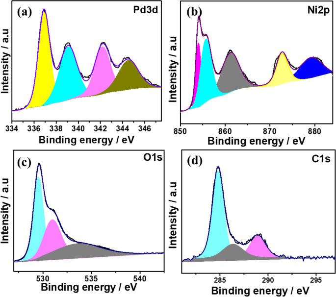
The digital state and chemical bonding of the PdO doped NiO composite was analyzed by utilizing XPS spectra. Determine 2 exhibits the XPS spectra of the PdO–NiO nanocomposite, the broad scan spectrum (Fig. SI. 1) of the PdO–NiO, which present the existence of the Pd (3d), Ni (2p), O (1s) and C(1s) components. The deconvoluted Pd 3d XPS spectrum in Fig. 2a exhibits two main peaks at a binding vitality of 336.4 and 342.2 eV corresponds to spin–orbit doublets of Pd 3d5/2 and Pd 3d3/2, respectively, which confirmed Pd2+ ions within the type of PdO within the PdO–NiO nanocomposite24,25. As well as, the satellite tv for pc peak of Pd species appeared at a binding vitality of 339.2 eV and 345.3 eV. In Ni 2p spectra (Fig. 2b), Ni 2p3/2 and Ni 2p3/2 spin–orbit doublets peaks have been noticed at 854.9 eV and 872.5 eV, which corresponded to Ni–O and Ni–OH, respectively and their corresponding satellite tv for pc peaks are situated at a binding vitality of 867.2 eV and 879.1 eV26,27. Moreover, O 1s spectra (Fig. 2c) present the 2 peaks at a binding vitality of 529.3 eV and 534.2 eV, which ascribed M–O and M–OH species. The obtained XPS spectra of the PdO–NiO nanocomposite are nicely related to the XRD and EDX evaluation.
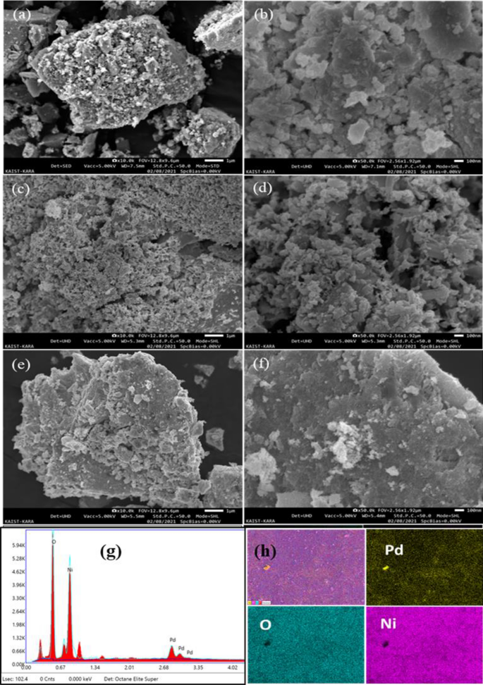
The morphology characteristic and elemental composition of the ready NiO, PdO and PdO–NiO nanocomposite was scrutinised by FE-SEM. Determine 3 exhibits the SEM morphology photographs of NiO, PdO and PdO–NiO nanocomposite at high and low magnification. Pure steel oxides NiO and PdO samples in Fig. 3a–d exhibits the porous crystalline morphology with excessive purity of the respective components. Alternatively, PdO doped NiO pattern in Fig. 3e,f exhibits the uniform, monodisperse, spherical crystalline morphology. Which confirms that PdO uniformly distributed with NiO. Therefore, the PdO doping enhances the floor space of the catalyst. As well as, the fundamental composition and elemental mapping was analysed for PdO–NiO pattern and proven in Fig. 3g,h. The EDX spectra and elemental mapping clearly confirms the presence of the Pd, Ni and O components within the composite with excessive purity.
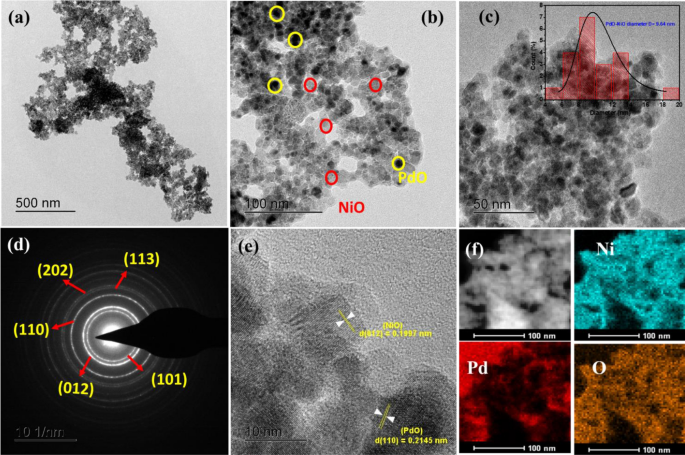
The detailed morphology and particle measurement distribution of the PdO–NiO NPs was measured by HR-TEM and the outcomes are introduced in Fig. 4. Determine 4a–c exhibits the everyday HRTEM photographs of the as-synthesized PdO–NiO nanocomposite. The obtained TEM photographs confirmed the uniform distribution of the spherical PdO–NiO NPs, which agrees with FESEM outcomes. In Fig. 4c (inset), the histogram reveals that shaped PdO–NiO nanoparticles are uniformly distributed with a median particle measurement of about 9.64 ± 2.1 nm, which is nicely related to XRD crystallite measurement. Moreover, the SAED sample was analyzed to know the crystallinity and the crystal high quality of the PdO–NiO nanoparticles are proven in Fig. 4d. The clear ring-like construction suggests the polycrystalline nature of PdO–NiO. The obtained diffraction rings d-spacing values are akin to the (101), (012), (110), (113) and (202) planes of the NiO nanoparticles. Determine 4e exhibits the lattice fringes of the PdO doped NiO nanoparticles. The fringes present the lattice planes for each steel oxides. The interplanar d-spacing worth of 0.1997 nm to correspond to the (012) airplane of the NiO part and the d-spacing worth of 0. 2145 nm to correspond to the (110) airplane of the PdO within the composite. Which is nicely correlated with the XRD d-spacing values. Elemental mapping in Fig. 4f exhibits the presence of Ni, Pd and O components with uniform distribution as related as SEM mapping. The morphology outcomes of the synthesized catalysts are nicely related to XRD, FTIR and XPS evaluation.
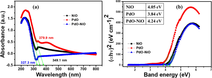
UV–Vis absorption spectra have been analyzed for the as-synthesized catalysts NiO, PdO and PdO–NiO and the respective outcomes are introduced in Fig. 5a. The absorption spectra present the strongest absorption maxima at 234.8 nm for all three catalysts. As well as, the attribute absorption band of NiO and PdO have been noticed at 338.2 nm and 422.1 nm respectively23, alternatively, no attribute absorption band was noticed for PdO–NiO pattern. Moreover, the bandgap vitality was calculated for 3 catalysts by utilizing the Schuster-Kubelka–Munk operate.
$$(alpha {textual content{h}}nu ),{textual content{n}} = {textual content{A}}({textual content{h}}nu – {textual content{Eg}})$$
(1)
The bandgap vitality (Eg) was achieved by extrapolating in opposition to the photon vitality and the obtained outcomes are proven in Fig. 5b. The calculated bandgap (Eg) of NiO, PdO and PdO–NiO are 4.05 eV, 3.84 eV and 4.24 eV, respectively25 (Fig. 5b, inset). The PdO doping with NiO will increase its bandgap worth. This implies that the PdO interface and NiO interface are carefully mixed within the composite. The obtained band hole worth of the catalysts is way increased than the reported bandgap vitality. The bandgap vitality is extremely depending on the particle’s measurement. The bandgap vitality will increase with reducing particle measurement, which confirmed that the synthesized catalysts are in nanoscale. The bandgap vitality (Eg) of the PdO–NiO catalyst is nicely related to FESEM and TEM outcomes.
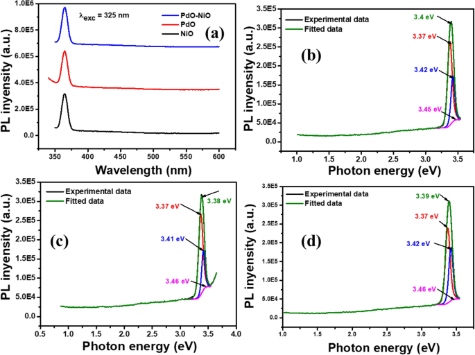
The photoluminescence (Pl) spectra of NiO, PdO and PdO–NiO supplies have been measured at 325 nm excitation wavelength and introduced in Fig. 6. Determine 6a exhibits the Pl spectra of the pure NiO, PdO and PdO–NiO nanocomposite. The blue/violet emission was noticed for all three samples at 364 nm because of the excitation of 3d8 electrons of Ni2+ ions from the conduction band to the valence band24. From Fig. 6a, it may be seen that the depth of the PdO–NiO nanocomposite is decrease than the pure NiO and PdO, which indicated the upper electron switch between the NiO and PdO, which is nicely correlated with electrochemical outcomes. The deconvoluted PL spectra of NiO, PdO and PdO–NiO supplies are proven in Fig. 6b–d; 4 peaks have been fitted for every pattern as proven in Fig. 6b–d. The UV emission at 364 nm (3.4 eV) corresponds to the close to band edge (NBE) excitation of NiO25. The obtained PL spectra confirmed that the PdO–NiO nanocomposite has extra conductivity than the pure steel oxides.
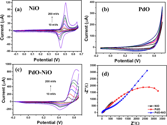
The electrode kinetics of the NiO, PdO and PdO–NiO modified GC electrode was explored in 1 M KOH at totally different scan fee variation at room temperature. As well as, the resistance of the aforesaid electrodes was monitored in impedance evaluation and proven in Fig. 7. Determine 7a, the NiO/GC present a pair of well-defined redox peaks at round 0.49 V and 0.44 V respectively, which akin to the reversible response between Ni2+ and Ni3+26. As well as, the redox peak currents linearly enhance with growing scan fee.
Determine 7b exhibits CV sample of PdO/GC electrode in 1 M KOH resolution, which sows poor peaks palladium oxide and palladium discount peak at 0.52 V and 0.53 V respectively because of the formation of oxyhydroxide on the electrode floor in fundamental medium. Whereas the PdO–NiO NPs modified GC electrode in Fig. 7c present well-defined Ni2+ and Ni3+ kinetics with eightfold increased peak present. Which confirms the Environment friendly electron switch between NiO and PdO within the composite. Which is nicely related to PL outcomes. Moreover, the impedance spectra have been achieved for 3 electrodes at mounted over potential (500 mV s−1) in 1 M KOH electrolyte and introduced in Fig. 7d. In Fig. 7d the Rct worth of the pure steel oxides NiO/GC and PdO/GC electrode have been obtained as 236 Ω and 2702 Ω respectively. Whereas the bimetal oxide PdO–NiO/GC present 425.7 Ω, which is decrease than the pure PdO. Because of the superior electron transformation between every steel oxide.
Catalytic discount of Azo compounds
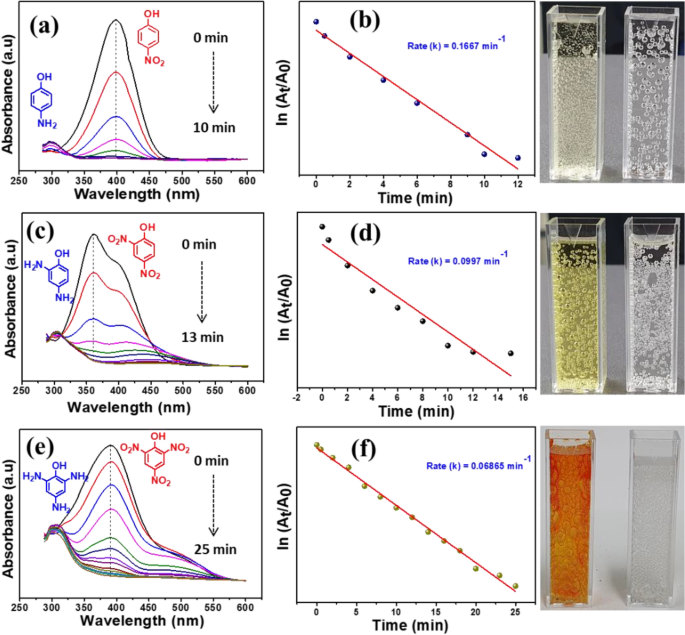
Azo compounds are extremely poisonous to the setting in addition to human beings. Particularly, nitrophenols are listed because the topmost hazardous chemical on this planet. Therefore the discount of nitrophenols positive factors probably the most consideration. Typically, the nitrophenol discount response is thermodynamically beneficial (E0 = − 0.76 V) at optimized circumstances, whereas the NaBH4 acts as a decreasing agent (E0 = − 1.33 V)27,28. Nonetheless, the discount fee is extended with out the catalyst because of the kinetic barrier between the decreasing agent and reactant. Therefore, the catalytic discount nitrophenols are a great way to transform to non-toxic aminophenol (AP) with the presence of NaBH4 as a decreasing agent. The discount response was simply monitored with a UV–Vis spectrometer. It’s identified that NaBH4 alone can’t scale back the nitrophenols into aminophenol’s. As proven in Fig. SI. 2 the contemporary nitrophenols (NP, DNP and TNP) absorption peak appeared at 300–370 nm respectively. When the addition of decreasing agent, the height was shifted to 402–450 nm28. As well as, the answer coloration was turned gentle yellow to deep yellow, because of the formation of corresponding nitrophenolate ions in fundamental resolution. Nonetheless, no discount was achieved over 2 h, indicating that nitrophenolate ions have been very secure with NaBH4. Moreover, the catalytic exercise of the NiO was explored with three nitrophenols and proven in Fig. SI. 3. The pure NiO reveals a poor catalytic discount of nitro compounds. In distinction, the PdO–NiO catalyst present glorious exercise on the nitrophenols, as proven in Fig. 8.
It may be seen that the nitrophenolate peak absorbance at 400 nm progressively decreases with response time, which confirmed that PdO–NiO promotes the electron and hydrogen switch between the reactant. Because of the increased energetic websites of the PdO–NiO. The current PdO–NiO catalyst completes the discount response of NP, DNP and TNP inside 10, 13, 25 min respectively. As well as, the aminophenol absorption peak appeared round 300 nm for all three nitrophenols. Alternatively, the deep yellow resolution turned colorless, indicating the formation of aminophenol. The speed fixed κapp for every nitrophenol was calculated from the plot of ln (At/Ao) Vs. time. The proposed PdO–NiO catalyst reveals glorious fee fixed 0.1667, 0.0997, 0.0686 min−1 for NP, DNP and TNT, respectively, which is the upper fee fixed than the beforehand reported catalyst (Desk SI. 2). Typically, the discount mechanism of nitrophenols to aminophenols follows many intermediate steps from nitro to nitroso after which to hydroxylamine and to remaining aminophenol. For these response required each electron switch and energetic hydrogen atoms. Right here, the BH4− ions produce the energetic hydrogen atoms on the floor of the catalyst and subsequently, the PdO–NiO catalyst enhances the electron switch. Consequently, the discount of NP might be effectively accelerated by the PdO–NiO catalyst. Moreover, comparability of the catalytic discount efficiency of nitrophenols with varies catalyst are proven in Desk SI. 2. The discount mechanism of the nitrophenols with PdO–NiO catalyst with NaBH4 as proven in Fig. SI. 6.
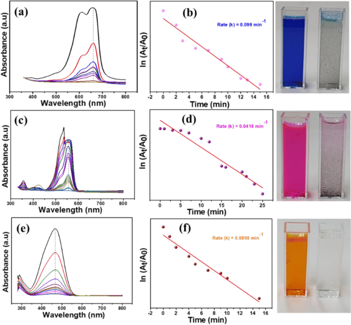
Moreover, the catalytic exercise of PdO–NiO composite was explored by the discount of natural azo dye compounds comparable to Methylene blue (MB), Rhodamine B (RhB) and Methyl orange (MO) with the addition of NaBH4 within the presence of PdO–NiO catalyst29. The discount of every dye was monitored at totally different absorption peaks, as proven in Fig. 9. The depth of every dye at respective wavelengths linearly lowered with time within the presence of the PdO–NiO. As well as, the discount fee κapp for every dye was calculated from the plot of ln (At/Ao) Vs. time. PdO–NiO catalyst exhibited glorious discount fee as 0.099, 0.0416 and 0.0896 min−1 for MB, RhB and MO, respectively, superior catalytic exercise than earlier studies. As well as, the azo dye resolution changed into colourless, indicating the whole discount happens within the presence of PdO–NiO. The pure NiO catalyst reveals poor discount exercise in direction of azo dyes with the presence of NaBH4 (Fig. SI. 4). Moreover, comparability of the catalytic discount efficiency azo dyes with varies catalyst are proven in Desk. SI. 3. Moreover, the discount of the combination of nitrophenols (NP, DNP and TNP) and azo dyes (MB, RhB and MO) was examined with PdO–NiO nanocomposite and obtained outcomes are proven in Fig. 10. Initially, the combination of azo compounds is shaped darkish resolution then quickly changed into a colourless and have become a clear resolution with the addition of PdO–NiO within the presence of NaBH430. The whole azo compounds discount was achieved inside 8 min. Therefore, the proposed PdO–NiO is a promising catalyst for wastewater remedy. As well as, the impact of the catalyst loading on the discount of combination of azo compounds have been studied with totally different loading quantity of PdO–NiO catalyst (3–10 mg) and proven within the Desk. SI. 4. Which present that the discount fee elevated with loading quantity of the catalyst. As well as, the catalytic discount efficiency of poisonous azo compounds by varied catalysts are proven in Desk. 2. PdO–NiO catalyst exhibit superior discount efficiency than the beforehand reported catalyst.
Moreover, the discount mechanism of azo dyes over PdO–NiO catalyst with decreasing agent proven in Fig. SI. 7.
After the whole discount response, the catalyst property was analyzed to know the steadiness of PdO–NiO. In Fig. 11a, FTIR spectra confirmed no noticeable modifications earlier than and after catalytic discount of combination discount. Moreover, the SEM picture (Fig. 11b) additionally confirmed no modifications within the morphology of the PdO–NiO. As well as, HR-TEM picture (Fig. 11c) was additionally analyzed to check the change within the particles measurement after catalytic discount, present no appreciable change within the particle measurement. Which proved that PdO–NiO is extremely secure within the discount circumstances.

