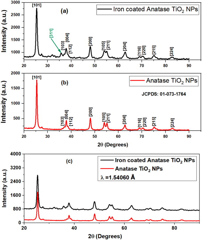
The samples have been subjected to Powder X-ray Diffraction utilizing a PANalytical X-Pert Professional X-ray diffractometer having Cu as anode materials and having Okayα1 as 1.54060 Å. Determine 2a depicts the XRD plot retrieved for Fe@TiO2 NPs and Fig. 2b exhibits the XRD sample obtained for naked TiO2 NPs. Determine 2b showcases anatase TiO2 NPs obtained on calcination at 400 °C. The height at 25.3764° confirms the [101] airplane of the anatase part. The whole sample matched properly with the JCPDS sample: 01-073-1764 confirming the peaks of anatase TiO2 NPs. Totally different planes acknowledged from the plot are [101] at 25.322°, [103] at 36.998°, [004] at 37.863°, [112] at 38.600°, [200] at 48.064°, [105] at 53.975°, [211] at 55.094°, [204] at 62.756°, [116] at 68.870°, [220] at 70.330°, [215] at 75.141° and [224] at 82.756°. Fig. 2a demonstrates the XRD peaks of Fe@TiO2 NPs and right here the iron-capping has been performed on the identical TiO2 NPs whose XRD obtained is proven in Fig. 2b. We clearly observe that each the patterns are similar and thus Fe@TiO2 NPs additionally possess the same anatase part, though the peaks are somewhat broadened. Whether or not the XRD sample of the core–shell NPs will match with that of the core NPs, this relies upon the thickness of the shell. The opportunity of Fe@TiO2 NPs having the same XRD sample could be attributed to the skinny layer of coating on the TiO2 NPs. The incident x-rays can simply penetrate the shell thickness of ~10 nm and beneath and thus virtually an identical sample is obtained. Furthermore, a cautious investigation signifies that one further peak has emerged at 35.631° which corresponds to airplane [311] of cubic Fe2O3 as denoted by JCPDS sample: 00-039-1346. That is justified because the coating of ferrous fumarate (Fe2+) was performed onto the TiO2 NPs. The crystallite dimension calculated utilizing Debye–Scherrer’s equation acknowledged the dimensions of ~12 nm for TiO2 NPs and ~17 nm for Fe@TiO2 NPs. Moreover, Fig. 2c depicts the superimposed XRD patterns obtained in each Fig. 2a, b. One can simply inform that the whole XRD plot of Fe@TiO2 NPs has been shifted up in depth. This can be a putting commentary that the core–shell Fe@TiO2 NPs retain the same XRD sample of the core TiO2 NPs. The essential thought of core–shell formulation is to have a uniform coating whereas nonetheless conserving the properties of the core. The layer shaped must not ever be too thick; in any other case, it’ll overshadow the options of the core25.
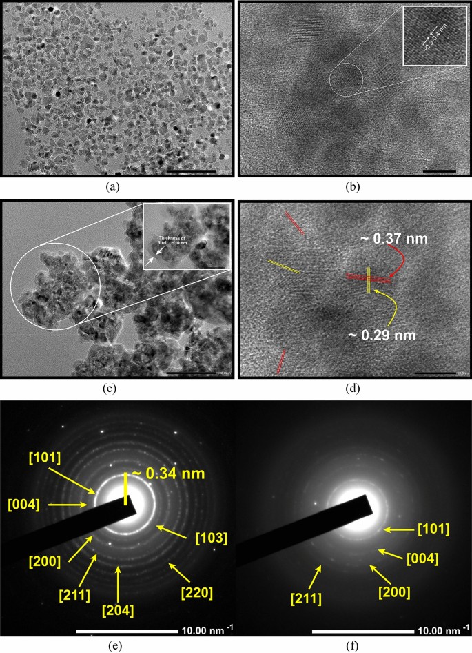
The Excessive-Decision Transmission Electron Microscopy (HR-TEM) pictures of TiO2 NPs are proven in Fig. 3a, b whereas Fig. 3c, d display the HR-TEM pictures of Fe@TiO2 NPs. Determine 3a exhibits that the typical dimension of the TiO2 NPs seem like ~10 nm. This goes together with the crystallite dimension calculated from the respective XRD plot. The particles are almost spherical and there’s a uniform dimension distribution. The interplanar distance lies between 0.3 and 0.4 nm, extra exactly 0.37 nm as noticed in Fig. 3b. The inset exhibits the magnified view of the area chosen to measure the interplanar distance. In Fig. 3c, we will clearly see the core–shell formulation of the NPs. Now, the NPs are not remoted, however agglomerated and an exterior layer/capping is distinctly seen. The darker parts point out the presence of component with decrease atomic quantity and right here relate to the core i.e. TiO2 NPs. The lighter parts point out the presence of a component with the next atomic quantity which is Fe2+ type of the iron on this case and corresponds to the shell. The inset shows the magnified view of the picture which has been used to measure the thickness of the layer. The size of the inset is 50 nm and with this, it’s deduced that the layer is at most 10 nm thick. In Fig. 3d, two completely different interatomic spacings of 0.37 nm and 0.29 nm could be seen overlapping in a lot of the areas. Determine 3e depicts the Chosen Space Electron Diffraction (SAED) sample for TiO2 NPs which matches properly with the corresponding XRD sample. Absolutely vibrant dots and distinct rings are clearly seen. This confirms the extremely crystalline nature of TiO2 NPs. Nonetheless, the SAED sample for Fe@TiO2 NPs in Fig. 3f consists of diffuse rings which signifies their much less crystalline nature. As soon as once more, this may be confirmed from the respective XRD sample in Fig. 2a. Thus, the findings obtained from XRD, HR-TEM and SAED evaluation are in good settlement with one another.
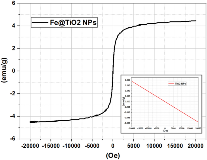
The Power Dispersive X-Ray Evaluation (known as EDX or EDS or generally EDAX) confirms the presence of parts Oxygen (O), Titanium (Ti) and Iron (Fe) in Fe@TiO2 NPs as proven in Fig. 4a. Equally, Fig. 4b depicts the presence of Oxygen (O) and Titanium (Ti) in TiO2 NPs. Desk 1 outlines the load share and atomic share of all the weather in each the samples. The Vibrating Pattern Magnetometer (VSM) research of each Fe@TiO2 NPs and TiO2 NPs are proven in Fig. 5. The inset of Fig. 5 illustrates the pure diamagnetic nature of anatase TiO2 NPs. The room temperature magnetism induced for the utilized magnetic area is a linear graph with a destructive slope indicating diamagnetism for TiO2 NPs. The plot of the diamagnetic conduct is much like the beforehand reported outcome26. Nonetheless, the VSM plot of TiO2 NPs capped with an iron complement capsule exhibits superparamagnetic conduct. Beketova et al.27 adorned TiO2 nanotubes with Fe3O4 NPs which additionally reported superparamagnetic conduct at room temperature. This can be a first in itself effort to magnetically modify the NPs utilizing commercially obtainable iron dietary supplements manufactured for human consumption. Thus, Fe@TiO2 NPs could be pushed to the goal websites with the assistance of exterior magnetic fields. Desk 2 summarizes the hysteresis parameters of Fe@TiO2 NPs obtained through VSM in opposition to Magnetization (emu) vs Utilized Magnetic Discipline (Oe).
For the drug launch research, we have now investigated the discharge conduct beneath completely different pH ranges. The next terminology has been used to point the samples in several pH options as proven in Desk 3.
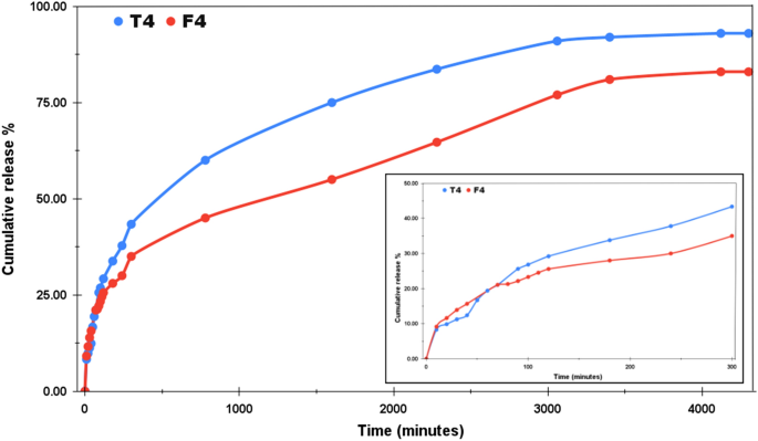
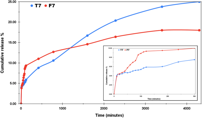
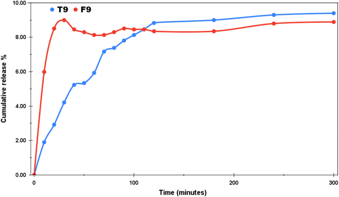
For each the acidic and the traditional mediums, the research was undertaken for 3 days. The selection of dialysis tube additionally impacts the sample of drug launch. Determine 6 represents the drug launch profile of T4 and F4 at pH 4.4 with cumulative launch share plotted alongside the y-axis and the time (in minutes) alongside the x-axis. The pH round carcinogenic tumors is often acidic, that’s why a lot of the chemo medicine are designed to ship sooner round acidic environments. Furthermore, the orally administered medicine attain first into the abdomen after consumption, the place the extremely acidic medium breaks it down into its elements from the place the molecules enter into the bloodstream. In our case, the preliminary launch within the first two hours was a lot sooner as in comparison with the overall research of three days. T4 and F4 launched upto 29.2% and 25.58% respectively on the finish of 120 min. T4 confirmed the cumulative launch of 60% in 14 h and eventually reached the utmost launch of 93% on the finish of our research. F4 displayed a fairly managed launch sample presenting approx. 50% cumulative launch after 26 h. The utmost launch % recorded for F4 on the finish of three days is 83%. The inset in Fig. 6 gives the drug launch sample for the primary 300 min. Thus, even beneath the acidic medium, each T4 and F4 confirmed a sustained launch sample and this attribute could be utilized for the managed supply of the drug. Likewise, Fig. 7 demonstrates the drug launch profile of T7 and F7 at pH 7.4. The pH of regular blood is mostly thought of to be between the vary 7.35–7.45. The NPs for use as drug carriers are usually anticipated to exhibit managed however sooner drug launch beneath acidic environments and fewer to no launch beneath regular pH. By this we will guarantee lesser drug wastage in the course of the circulation and simpler drug launch on the goal areas. Below regular pH, we will see that within the span of three days, the utmost cumulative launch share was 25% and 18% for T7 and F7 respectively. No additional launch was noticed and F7 has outperformed T7 within the context of least drug launch within the regular pH. The inset in Fig. 7 exhibits the preliminary launch sample within the first 300 min. T7 offered 5.87% launch and F7 confirmed 9.34% cumulative launch within the preliminary 300 min. Moreover, the samples have been additionally examined beneath the essential medium of pH 9 and as anticipated, an total launch of 9.4% was noticed for T9 and eight.9% was noticed for F9 as proven in Fig. 8. This research was solely stretched for 300 min as no considerable launch was seen after 120 min. This research confirms that the drug launch charge of each TiO2 NPs and Fe@TiO2 NPs is pH-dependent. The utmost launch has been detected within the acidic medium and the least launch within the primary medium. Thus, each sorts of NPs are appropriate candidates for use as drug-delivery brokers. Fe@TiO2 NPs exhibited a extra managed drug-release conduct and due to this fact, the maximal launch attained by them is lower than TiO2 NPs beneath all of the pH ranges.
In-vitro research performed by Kadivar et al.28 means that 99.56% of the Imatinib is launched inside 20 min in 0.1 N HCl resolution. Compared to this commentary, each the NPs synthesized by us display a lot managed and sustained launch of the drug (Imatinib) which may be very useful for an efficient chemotherapeutic therapy. If the drug is retained for an extended time, this may decrease the drug dosages required which might additional management the side-effects. The outcomes of the in-vitro research revealed the attainment of most drug launch beneath an acidic setting by these NPs, suggesting that they endure comparatively sooner decomposition thereby selling sooner launch at low pH degree than at impartial pH degree. CaCO3 NPs display comparable pH-responsive behaviour and have been extensively studied for drug supply functions. They exhibit low toxicity, gradual biodegradability and when functionalized, they not solely increase the effectiveness of drug-delivery, but additionally improve drug-loading, tumor-targeting and thermodynamic stability29. Thus, NPs should be improved for showcasing a number of advantages.
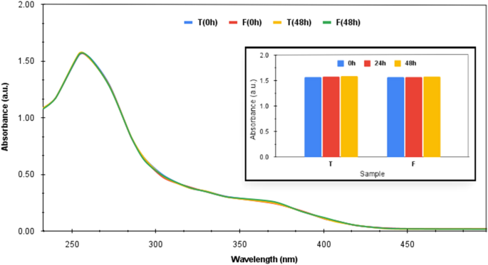
Determine 9 showcases the UV–seen plots for each the NPs taken at 0 h i.e. initially of the research and at 48 h (on the finish of the research), respectively. ‘T’ represents the TiO2 NPs and ‘F’ represents the Fe@TiO2 NPs. The inset of Fig. 9 exhibits the comparability of the absorbance of the NBT resolution recorded at 254 nm over three completely different intervals of time i.e. 0 h, 24 h and 48 h respectively within the presence of each the NPs individually. It’s observable that there was no important lower within the absorbance of the attribute peak at 254 nm over the time period. The plots of TiO2 NPs-NBT resolution and Fe@TiO2 NPs-NBT resolution taken over completely different instants of time virtually overlapped. Furthermore, no color change was detected by observing the samples with bare eye. Thus, the ROS detection take a look at performed by us for each the NPs validate that no ROS is generated by these NPs beneath human physiological situations which confirms their biosafety. Basante-Romo et al.30 modified TiO2 NPs with functionalized multiwalled carbon nanotubes and evaluated them for his or her degree of toxicity. The toxicity research have been carried out on feminine albino rats and no opposed results have been noticed after the 10-days research. No mortality was produced. All of the scientific indicators have been regular suggesting that no adjustments occurred within the cells or tissues exhibiting regular structure of the organs. Thus, the modification of TiO2 NPs diminishes the possibilities of them being damaging. The coating of those NPs with a biocompatible materials and different types of floor modifications guarantee that these TiO2 NPs will not be displaying their ROS technology means sans irradiation. Since these NPs aren’t irradiated when used for drug-delivery functions, the possibilities of them leading to deadly ROS are very low.
Future perspective
The utilization of NPs as drug-delivery brokers belong to new-era medical methods. A number of analysis is occurring to make sure the suitability of those NPs for the mentioned function. This very thought remains to be in its infancy and it has to efficiently cross a loads of pre-clinical and scientific trials to be really in a position for a licensed medical utilization. Speaking about guaranteeing focused drug supply of chemotherapeutics, emphasis must be laid on quite a lot of components together with the mitochondrial response, the oxidative stress, the receptors current, any kind of inherent or induced drug resistant response of the mobile elements and lots of extra. Moreover, any type of toxicity provided by the NPs must also be handled nice care. The toxicity of the NPs doesn’t lie solely within the formation of ROS, however the dimension and form of the NPs are additionally the components of grave concern. The NPs must be optimally synthesized to have optimum dimension and form which provide least accumulation within the tissues and organs. Totally different methods to functionalize the NPs, to change their floor properties, to optimize their dimension and form, to make sure ample quantity of drug loading onto them, to enhance their antioxidant traits and to make them biocompatible, must be devised. Thus, ample steps must be taken to make sure the biosafety of the NPs for use as drug-carriers and to additional enhance their drug-delivery response. As soon as passable outcomes are achieved from in-vitro research, solely then extra systematic approaches could be explored to information in-vivo analysis and higher correlate the properties of nanoparticles with their organic results.

